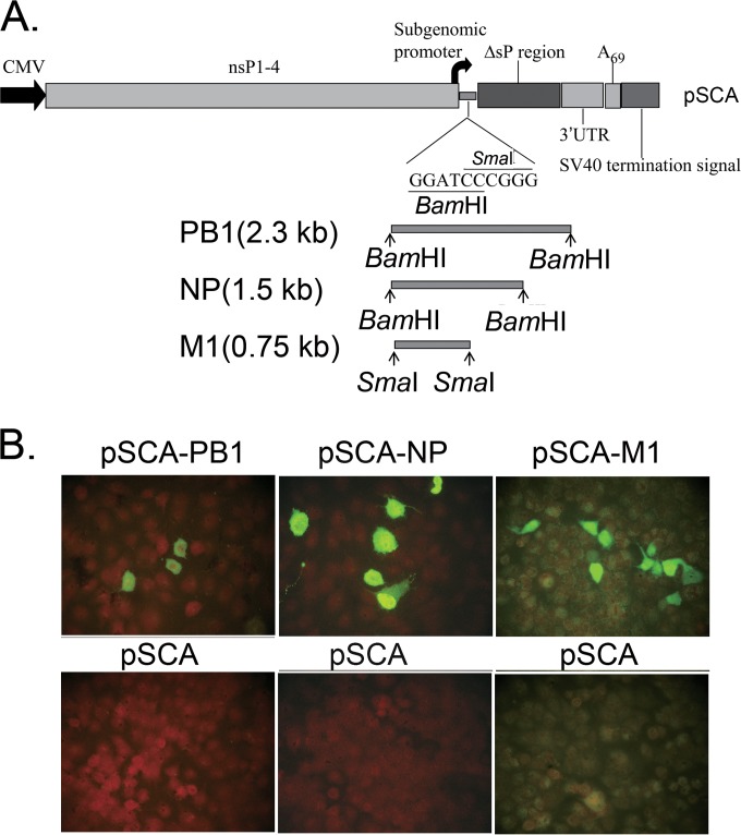
FIG 1.
Genetic organization of DNA vaccines based on PB1, NP, and M1 in pSCA vector and the resulting protein expression in MDCK cells transfected with recombinant plasmids. (A) Schematic diagram of the genetic organization of the pSCA DNA vector (top). The bottom shows the cDNA fragments of influenza viruses BJ95 PB1, NP, and M1 flanked by BamHI, BamHI, and SmaI restriction sites, respectively. CMV, cytomegalovirus. (B) Indirect immunofluorescence showing the expression of influenza PB1, NP, and M1 in MDCK cells transfected with pSCA-PB1 (left), pSCA-NP (middle), and pSCA-M1 (right) recombinant plasmids (top) stained with the polyclonal or monoclonal antibodies (Abs) indicated in Materials and Methods. The bottom images show the results of MDCK cells mock transfected with pSCA plasmid and detected using the same Abs as in the top images.

