
Figure 3.
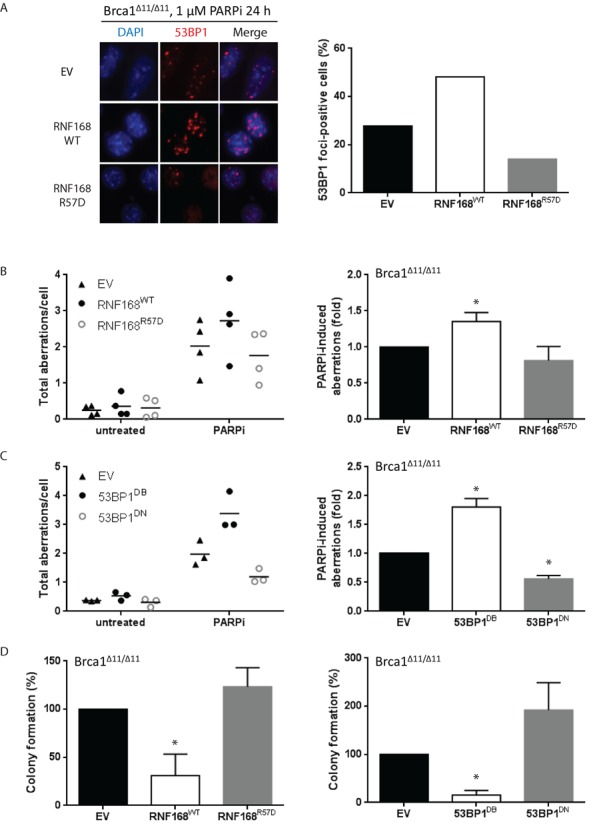
Overexpression of RNF168 or 53BP1 exacerbates PARP inhibitor-induced genomic instability and cytotoxicity. (A) BRCA1Δ11/Δ11 cells were treated with 1 μM PARPi for 24 h and processed for standard immunofluorescence. Left panel: cells were stained for 53BP1 (red) and imaged at 63× magnification. A representative experiment is shown. Right panel: the percentage of cells that contain >5 foci of 53BP1 from two independent experiments. At least 200 cells were scored for each sample and treatment condition. (B) BRCA1Δ11/Δ11 MEFs stably transduced with retroviral vectors encoding RNF168WT or RNF168R57D were treated with 1 μM PARPi (24 h) and harvested for preparation of metaphase spreads. Left panel: dot plots indicating the total amount of aberrations per cell in four independent experiments. At least 100 metaphases were analyzed for each condition. Right panel: histograms depicting PARPi-induced chromosomal aberration load relative to empty vector-transduced cells for the same experiments as shown in the corresponding left panels. (C) Similar to (B), except in BRCA1Δ11/Δ11 MEFs stably transduced with retroviral vectors encoding 53BP1DB or 53BP1DN. (D) Cells were treated with 1 μM PARPi for 24 h and then incubated in drug-free medium to allow formation of colonies. After 9 days, culture dishes were stained with crystal violet and colonies containing >50 cells were counted. Results are mean ± SD of three independent experiments. For (A)–(D), Statistical significance was determined with two-tailed unpaired Student's t-test; *, P < 0.05 compared to empty vector-transduced cells.