
Figure 4.
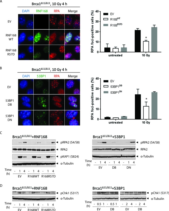
RNF168 and 53BP1 block RPA foci formation and RPA2 phosphorylation. (A) BRCA1Δ11/Δ11 MEFs stably transduced with retroviral vectors encoding RNF168WT or RNF168R57D were irradiated with 10 Gy and fixed 4 h later. Samples were processed for standard immunofluorescence. Left panel: cells were co-stained for RNF168 (green) and RPA2 (red) and imaged at 63× magnification. Note that the polyclonal anti-RNF168 antibody used in this study recognizes only the exogenously expressed human RNF168. A representative experiment is shown. Right panel: the percentage of cells that contain >10 RPA2 foci. (B) BRCA1Δ11/Δ11 MEFs stably transduced with retroviral vectors encoding 53BP1DB or 53BP1DN were irradiated and processed for standard immunofluorescence as in (A). Left panel: cells were co-stained for 53BP1 (green) and RPA2 (red) and imaged at 63x magnification. A representative experiment is shown. Right panel: the percentage of cells that contain >10 RPA2 foci. The right panels in A and B show mean ± SD of three independent experiments. At least 200 cells were scored for each sample and treatment condition. (C) Similar to (A), except cells were harvested at the indicated post-irradiation time points for western blot analysis. (D) Similar to (B), except cells were harvested at the indicated post-irradiation time points for western blot analysis. A representative blot is shown in (C) and (D). Experiments were repeated three times. For (A) and (B), statistical significance was determined with two-tailed unpaired Student's t-test; *, P < 0.05 compared to empty vector-transduced cells.