
Figure 2.
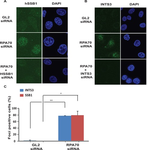
INTS3 and hSSB1 form sub-nuclear foci after RP70 depletion. (A and B) HeLa cells transfected on three consecutive days with GL2 or RPA70 siRNA in combination with HSSB1 or INTS3 siRNA as indicated were visualized for hSSB1 and INTS3 foci by immunofluorescence with rabbit anti-hSSB1 and goat anti-INTS3 antibodies respectively. Right panel displays the DAPI staining for each sample. Co-depletion of hSSB1 (A) or INTS3 (B) along with RPA70 confirms that the immunofluorescence signal is from the respective proteins. (C) Quantification of INTS3 and hSSB1 foci observed in the experiments described in parts A and B. Cells from GL2 or RPA70 siRNA transfected samples were scored for INTS3 and hSSB1 foci and are expressed as a percentage of total cells from each group. Data are represented as the mean ± SE. P-values were calculated using two-tailed t-test which displays that RPA70 siRNA transfected samples are significantly different from control GL2 siRNA transfected samples (*P-value < 0.05; **P-value < 0.005).