
Figure 1.
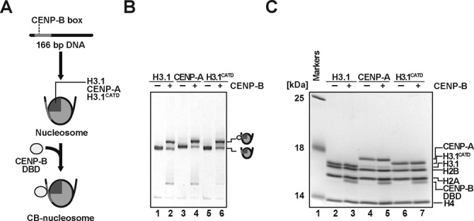
CENP-B binds to the CENP-A and H3.1 nucleosomes. (A) Schematic representation of CENP-B DBD binding to nucleosomes. (B) Electrophoretic mobility shift assay. The H3.1, CENP-A and H3.1CATD nucleosomes (lanes 1, 3 and 5, respectively) and those complexed with the CENP-B DBD (lanes 2, 4 and 6, respectively) were analyzed by non-denaturing 6% polyacrylamide gel electrophoresis with ethidium bromide staining. (C) Protein contents of the H3.1, CENP-A and H3.1CATD nucleosomes (lanes 2, 4 and 6, respectively) and those complexed with the CENP-B DBD (lanes 3, 5 and 7, respectively), analyzed by SDS-15% polyacrylamide gel electrophoresis with Coomassie Brilliant Blue staining.