
Figure 4.
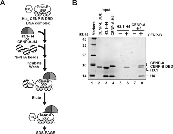
The CENP-B DBD specifically binds to the CENP-A-H4 complex. (A) Schematic representation of the pull-down assay with the His6-tagged CENP-B DBD and the CENP-A-H4 or H3.1-H4 complex. (B) The His6-tagged CENP-B DBD (50 nM) complexed with a 21 base-pair DNA was incubated with the CENP-A-H4 or H3.1-H4 complex (50 nM). The proteins pulled down with the Ni-NTA agarose beads were analyzed by 16% SDS-PAGE with Coomassie Brilliant Blue staining. Lane 1 indicates the molecular mass markers. Lane 2: His6-tagged CENP-B DBD (50% of input). Lane 3: the H3.1-H4 complex (20% of input). Lane 4: the CENP-A-H4 complex (20% of input). Lanes 5 and 6 represent the pull-down experiments with the H3.1-H4 complex, in the absence and presence of the His6-tagged CENP-B DBD, respectively. Lanes 7 and 8 represent the pull-down experiments with the CENP-A-H4 complex, in the absence and presence of the His6-tagged CENP-B DBD, respectively.