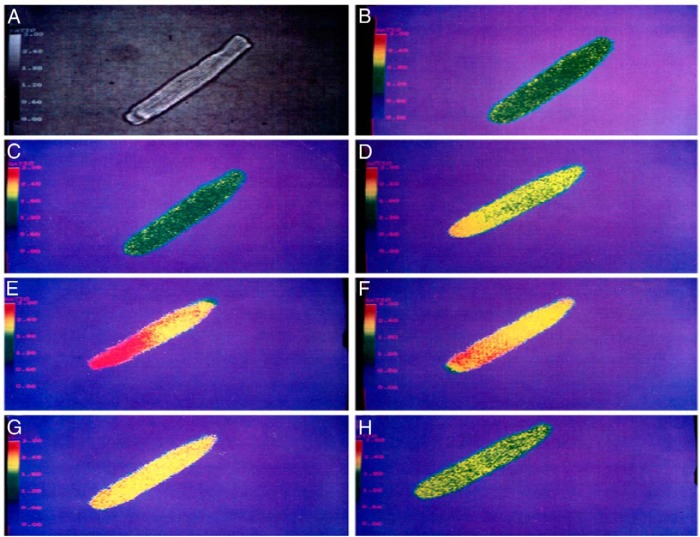
Figure 2.
Nonlytic transient MAC triggers a large increase of intracellular Ca++ concentration ([Ca++]i). The figure shows pseudocolor images of isolated adult rat ventricular muscle cell loaded with fura-2, a fluorescent calcium-indicator dye. Increasing emission intensities were associated with increasing [Ca++]i, coded in a pseudocolor spectrum from blue (low Ca++) to red (high Ca++). A, Reference, digitalized phase-contrast image. Before adding complement factors, the cell shows low-emission intensity (yellow-green) (B) that does not significantly change after the addition of C5b6 plus C7 and C8 (C). After addition of C9, the cell showed a marked emission increase (D and E), representing an increase in [Ca++]i that lasted approximately 1 minute. After an additional 3 minutes, [Ca++]i decreased to near baseline levels (F–H). [Reproduced from H.J. Berger et al. Activated complement directly modifies the performance of isolated heart muscle cells from guinea pig and rat. Am J Physiol. 1993;265:H267–H272 (36). J.A. Halperin was an author, no permission needed by The American Journal of Physiology to reproduce.]

