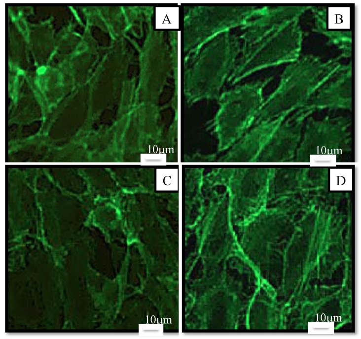
Figure 6.
Morphology of human endothelial cells observed under fluorescence microscope after 48 h in culture medium (control (A)) or in samples at a concentration of 10 µM in culture medium (AstaS (B), AstaP (C) and AstaCO2 (D). Cells were stained with Alexa Fluor-conjugated phalloidin for detection of actin filaments. Scale bar: 10 μm.

