Figure 6.
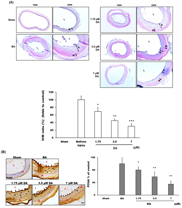
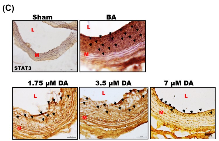
Dihydroaustrasulfone alcohol prevents balloon angioplasty-induced neointima formation. (A) The arterial sections were stained with hematoxylin-eosin (H&E) to observe the thickness changes of vessel wall. The images were acquired by microscopy at 100–200× magnification. The manifestation of vascular restenosis was presented as the ratio of neointima-to-media area (N/M ratio); (B) The distribution and expression of proliferating cell nuclear antigen (PCNA) protein were detected with immunohistochemistry analysis, and the images were acquired by microscopy at 400× magnification; (C) The distribution and expression of signal transducer and activator of transcription 3 (STAT3) protein were detected with immunohistochemistry analysis, and the images were acquired by microscopy at 400× magnification. L, lumen; N, neointima layer; M, media layer. The black arrow indicated the position with the expression of detected proteins.* p < 0.05, ** p < 0.01 and *** p < 0.001 compared with balloon angioplasty (BA) group, respectively.


