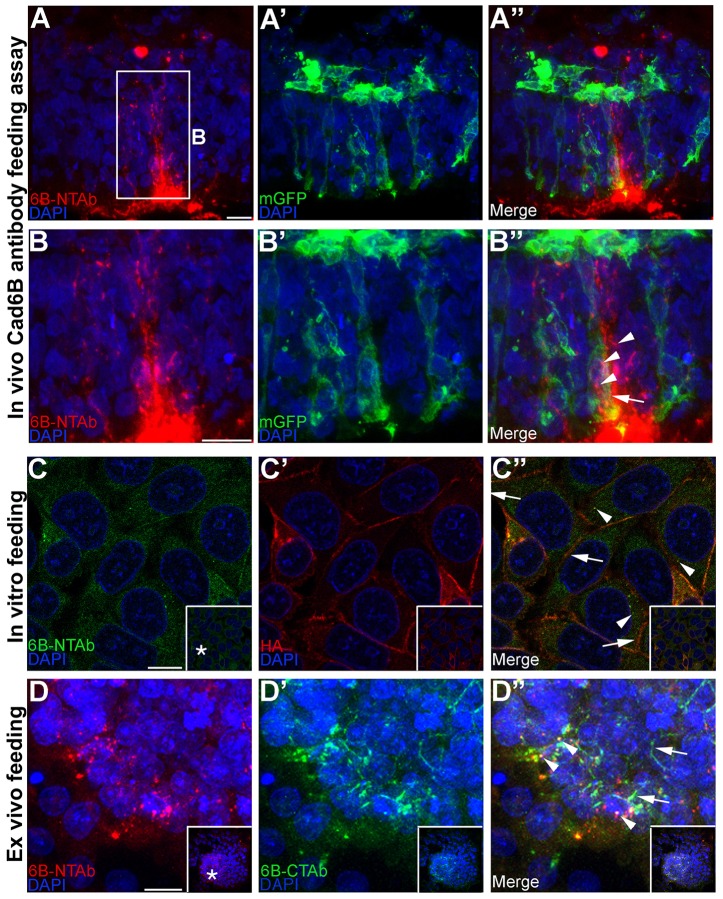
Fig. 2.
Cad6B puncta are endocytic rather than exocytic in nature. (A–A″) Cad6B antibody recognizing the Cad6B extracellular domain (NT-6B) was introduced into the lumen of embryos electroporated with membrane-associated GFP, incubated to allow for EMT, fixed and immunostained with the NT-6B antibody (red) and GFP (green). (B–B″) High magnification image of the boxed region in A. Internalized NT-6B antibody–Cad6B complexes are seen in the cytoplasm (B″, arrowheads) within the confines of GFP-expressing plasma membrane (B″, arrow) in a delaminating neural crest cell. The NT-6B antibody was added to medium of FlpIn-wtCad6B cells (C–C″) and dorsal neural fold explants (D–D″), followed by incubation to allow for EMT, a low pH buffer wash, fixation and immunostaining for NT-6B (C,D) and HA (C′) or CT-6B (D′). Internalized NT-6B antibody–Cad6B complexes localize to the cytoplasm (C″,D″, arrowheads), and Cad6B is still observed on the plasma membrane (C″,D″, arrows). All images except C–C″ represent the 3D composite of several Z-stack images acquired with the confocal. Inset boxes in C–D″ show the original image, with the asterisks in C and D indicating the location of the higher magnification field in the main panels. Scale bars: 10 µm.

