Fig. 3.
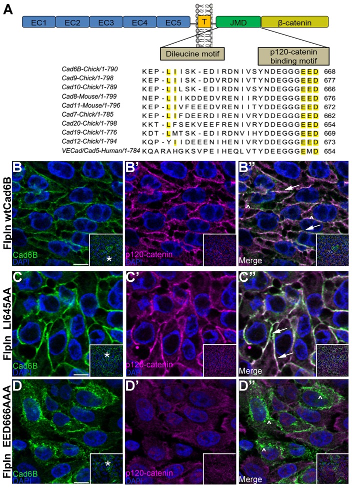
Cad6B possesses putative endocytic motifs in its cytoplasmic domain. (A) A portion of the alignment of the Cad6B juxtamembrane domain with several type II cadherins. The dileucine and putative p120-catenin binding motifs are highlighted (yellow). EC, Extracellular domain; T, transmembrane domain; JMD, juxtamembrane domain. FlpIn cells expressing wtCad6B (B–B″), LI645AA (C–C″) and EED666AAA (D–D″) were fixed and immunostained for Cad6B (green) and p120-catenin (purple). Panels represent single confocal plane images. Arrows point to membrane-bound Cad6B and p120-catenin, and carets indicate Cad6B-positive, p120-catenin-negative cytoplasmic puncta. Inset boxes in B–D″ show the original image, with the asterisks in B,C,D indicating the location of the higher magnification field in the main panels. Scale bars: 10 µm.

