Fig. 7.
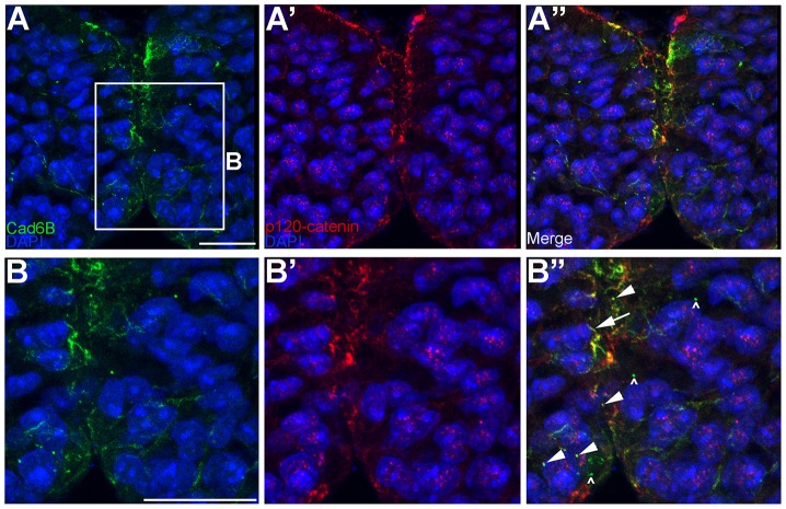
Cad6B partially colocalizes with p120-catenin in cytosolic puncta in vivo. (A–A″) Embryos in which neural crest cells are actively undergoing EMT were fixed and immunostained for Cad6B (green) and p120-catenin (red). (B–B″) A higher magnification view of the boxed region in A. Panels represent the 3D composite of several Z-stack images acquired with the confocal. Arrowheads in B″ point to colocalized Cad6B and p120-catenin in the cytosol, the arrow shows colocalized Cad6B and p120-catenin at the membrane and carets show Cad6B-positive, p120-catenin-negative puncta. Scale bars: 10 µm.

