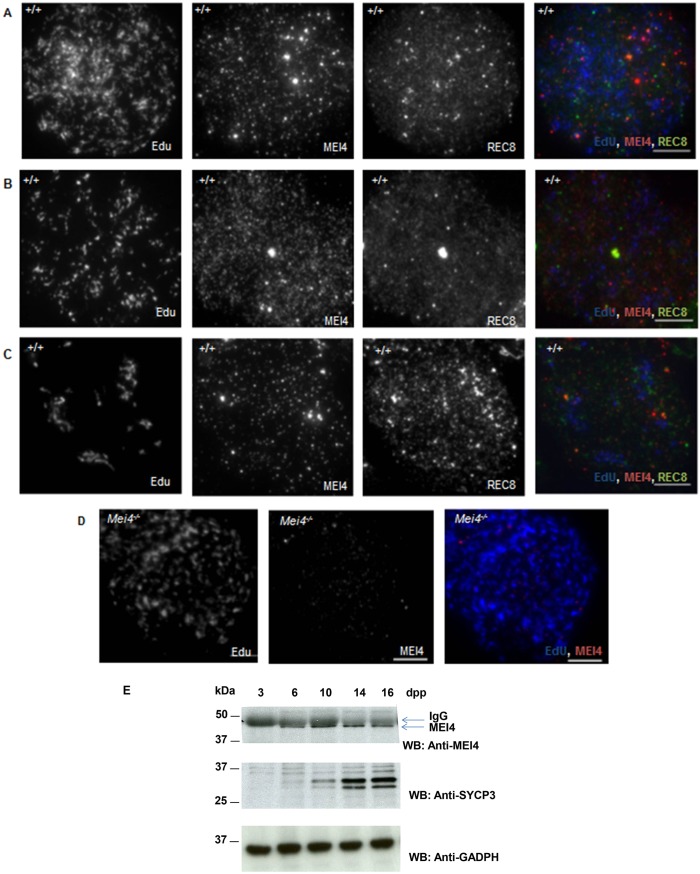
Fig. 2.
MEI4 and REC8 expression in preleptotene cells. MEI4 localization on meiotic chromosome spreads prepared after in vitro EdU labeling for 1 h of cells from wild-type (A–C) and Mei4−/− testes (D). In wild-type spreads, REC8 localization was concomitantly assessed. Preleptotene nuclei are in early (A), middle (B) and late (C) S phase, based on EdU localization relative to heterochromatin identified by intense DAPI staining (not shown). (E) MEI4 immunoprecipitation from protein extracts of juvenile testes at different times during the first wave of spermatogenesis with a non-crosslinked anti-MEI4 antibody. SYCP3 and GAPDH are used as controls. dpp, day postpartum. Scale bars: 10 µm.

