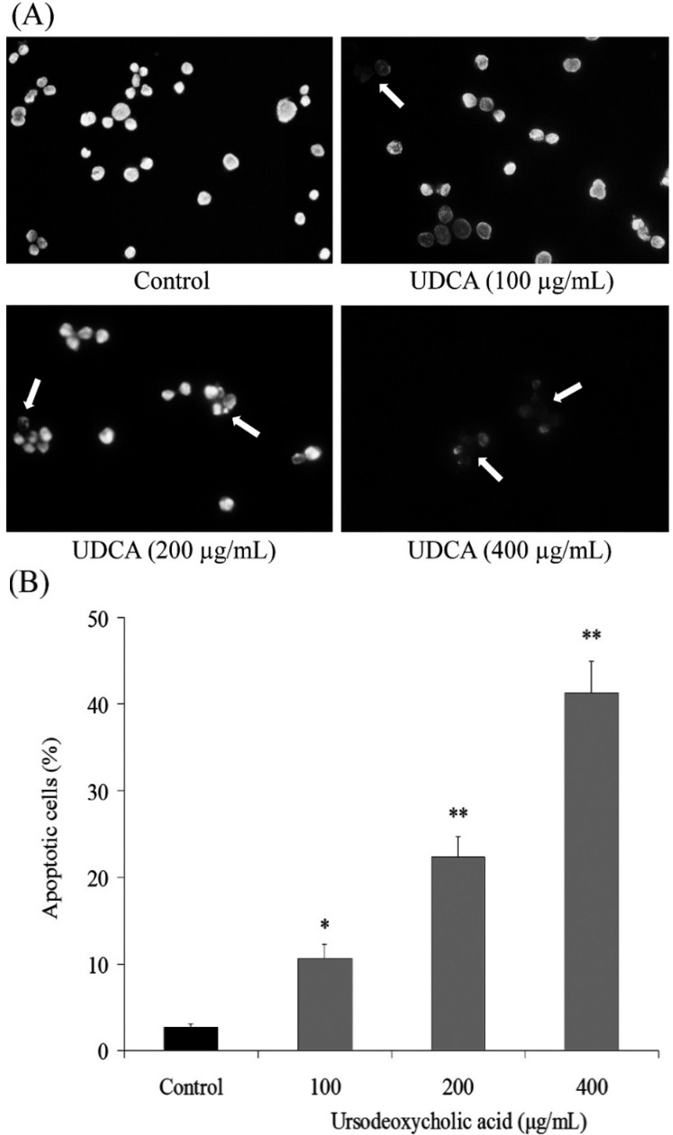
Figure 2.
Exposure of human oral squamous carcinoma HSC-3 cells to ursodeoxycholic acid (UDCA) induced apoptosis. (A) Appearance of apoptotic bodies in HSC-3 cells treated with UDCA for 48 h (200×); and (B) treatment with UDCA for 48 h increased the number of apoptotic cells as measured by flow cytometry. The profile represents an increased sub-G1 population (apoptotic cells). * Mean values are significantly different from control (p < 0.05) and ** mean values are significantly different from control (p < 0.01).

