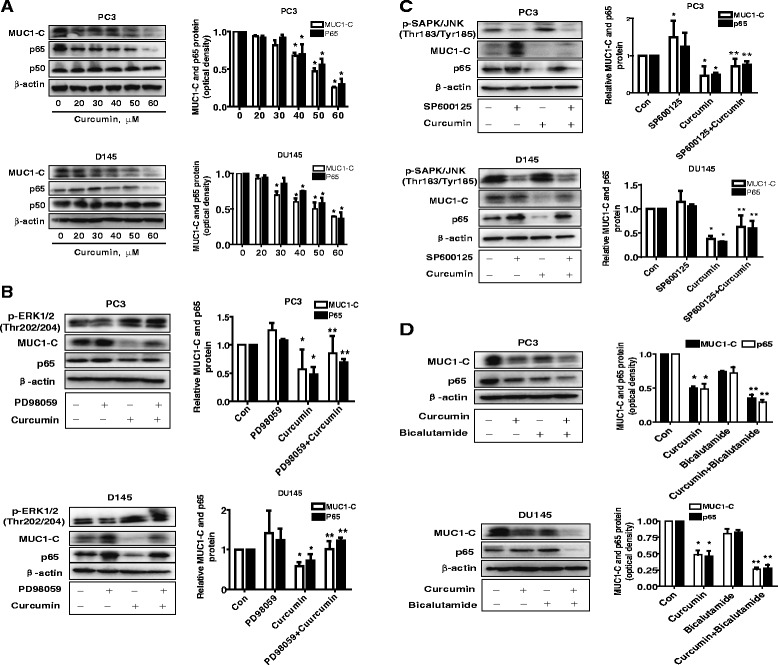
Fig. 3.

The effect of curcumin and bicalutamide on protein expression of p65 and MUC1-C through activation of ERK/12 and SAPK/JNK. a PC3 and DU145 cells ells were exposed to increased concentration of curcumin for 24 h. Afterwards, the expression of p65 and MUC1-C proteins was detected by Western blot. b-c PC3 and DU145 cells were treated with PD98059 (10 μM) and SP600125 (20 μM) for 2 h before exposure of the cells to curcumin (40 μM) for an additional 24 h. Afterwards, the expression of p65 and MUC1-C protein were detected by Western blot using antibodies against p65 and MUC1-C. The bar graphs represent the mean ± SD of p65 or MUC1-C /GAPDH of three independent experiments. d, PC3 and DU145 cells were treated with curcumin (40 μM) and bicalutamide (30 μM) for 24 h. Afterwards, the expression of p65 and MUC1-C protein were detected by Western blot using antibodies against p65 and MUC1-C. Values in bar graphs were given as the mean ± SD from three independent experiments performed in triplicate. *indicates significant difference as compared to the untreated control group (P < 0.05). **Indicates significant difference from curcumin treated alone (P < 0.05)
