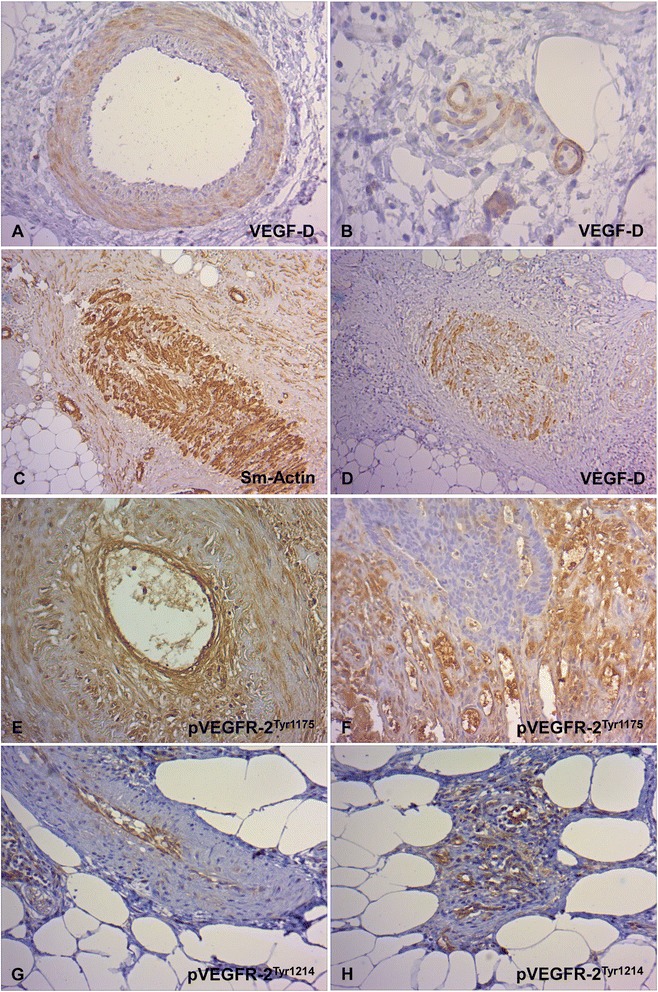
Fig. 3.

Immunohistochemical staining of the VEGF-D and pVEGFR-1 in the vasculature of CC tissue. a,b Characteristic vascular VEGF-D expression. VEGF-D-positive macrovascular vessels with immunoreactivity in smooth muscle cells of the media layer and occasionally in endothelial cells (a, x 200) and VEGF-D-positive capillaries with endothelial immunoreactivity (b, x 400). Altered macrovascular vessels with discontinuous, hypoplastic smooth muscle cell layer (c, Sm-Actin., x 100) and VEGF-D immunopositivity (d, x 100). e,f Characteristic endothelial VEGFR-2Tyr1175 expression in macrovessels (e, x 200) and capillaries (f, x200). g,h Characteristic endothelial VEGFR-2Tyr1214 expression in macrovessels (g, x 200) and capillaries (h, x 200)
