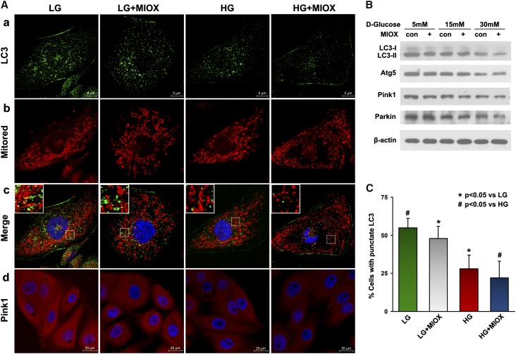
Figure 3.
Accelerating effect of MIOX on autophagy, mitophagy, and their associated protein expression under HG ambience. (A, a–c) Fluorescent punctate LC3 staining (green) was reduced in MIOX overexpressing cells in both LG and HG ambience, and this was associated with altered Mitored staining (red) and decreased LC3 and Mitored merged dots (yellow), indicating increased mitochondrial fission and decreased autophagy and mitophagy. (B) Western blot analyses revealed a reduced expression of LC3-II and Atg5 in a dose-dependent manner in cells subjected to HG treatment, and their expression was further attenuated by MIOX overexpression, whereas Atg5 expression was not obviously affected. (C) Quantification also indicated reduced cell percentage with punctate LC3 staining. (A, d and B) The expression of mitophagy-related proteins, Pink1 and Parkin, was also reduced under HG ambience, and their expression was further suppressed by MIOX overexpression when subjected to HG. Con, control.

