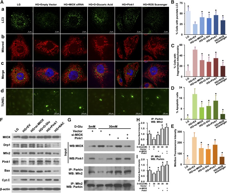
Figure 5.
Attenuation of mitochondrial fragmentation, ROS, and apoptosis but increased autophagy and mitophagy after inhibition of MIOX under HG ambience. (A, a–c, B, C, and F) Under HG, cells transfected with MIOX siRNA or incubated with d-glucaric acid had increased expression of punctate LC3 and Pink1, indicative of increased autophagy/mitophagy, as well as decreased mitochondrial fragmentation and altered expression of mitochondrial shaping proteins. (A, d and D–F) This was accompanied with decreased mitochondrial ROS production, reduced expression of Bax and Cytochrome C, and apoptosis compared with empty vector-transfected cells treated with HG. MIOX may modulate mitochondrial quality control through two different mechanisms (i.e., Pink1 pathway and MIOX-induced oxidative stress). (A, columns 5 and 6 and F) Pink1 pcDNA-transfected cells had reduced mitochondrial fragmentation and Drp1 levels but increased Mfn2 expression together with increased autophagy and mitophagy and decreased ROS generation and apoptosis. Similarly, the cells pretreated with ROS scavenger N-acetylcysteine (NAC) and subjected to HG showed partial recovery of mitochondrial integrity and reduced apoptosis. (E and F) These changes were similar to those seen with MIOX inhibition, whereas MIOX has been shown to induce ROS and inhibit Pink1 expression. (G–I) The coimmunoprecipitation experiments indicated that, in tubular cells, HG inhibited the Parkin–Mfn2 interaction, whereas knockdown of MIOX upregulated Pink1 expression and increased the Parkin–Mfn2 interaction. The overexpression of Pink1 had similar and more prominent effects on such interactions. Cyt.C, Cytochrome C; D-Glu, D-glucose; IP, immunoprecipitation; WB, Western blot. *P<0.05 compared with HG plus the empty vector group.

