Figure 8.
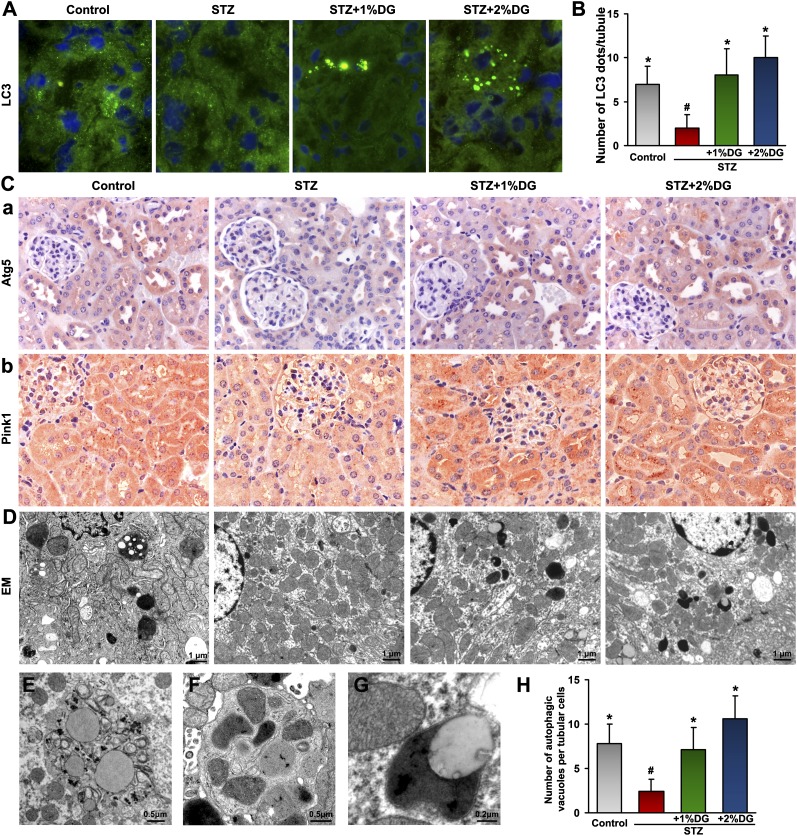
Restorative effect of d-glucarate on autophagy and mitophagy in STZ-induced diabetic mice. (A) The basal level of autophagy, indicated by punctate LC3 staining, was seen in the renal tubules of the control group, which disappeared in the tubules of the STZ group, whereas they were restored after d-glucarate treatment. (B) Morphometric analyses confirmed these observations. (C) The expressions of Atg5 and Pink1 exhibited parallel reduction and restoration in the tubules of mice from four groups. (D and H) By electron microscopy, autophagic vacuoles seen in the control group were greatly reduced in proximal tubular cells in the STZ group, whereas they were restored in tubules from mice treated with d-glucarate. (E) Higher magnification electron micrographs revealed a typical morphology of an autophagosome containing fragmented cellular organelles, (F) a mitophagosome, and (G) an autolysosome in tubular cells after d-glucarate treatment. DG, d-glucarate; EM, electron microscopy. *P<0.05 compared with the STZ group; #P<0.05 compared with the control group.

