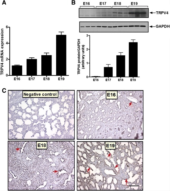
Fig. 1.

TRPV4 expression increases with gestation. a Fetal lung tissue was collected at different times in gestation, as shown. RNA was isolated, reversed-transcribed, and the cDNA products for TRPV4 were analyzed by quantitative RT-PCR. N = 3. b Fetal lung tissue was collected at different times in gestation and proteins extracted to assess TRPV4 expression by Western blot. The upper panel is a representative blot. Results were normalized to GAPDH to control for protein loading. N = 3. c Fetal lung tissue from different times in gestation was fixed in formalin. Sections were process by immunohistochemistry using rabbit anti-TRPV4 polyclonal antibody, stained with diaminobenzidine and counterstained with hematoxylin. Immunohistochemistry pictures show distribution of TRPV4 during fetal lung development (arrows). At E16 (pseudoglandular stage), TRPV4 was minimally expressed in the respiratory bronchioles. Later in gestation, TRPV4 immunostaining was more apparent in the distal epithelium. Bar, 20 μm
