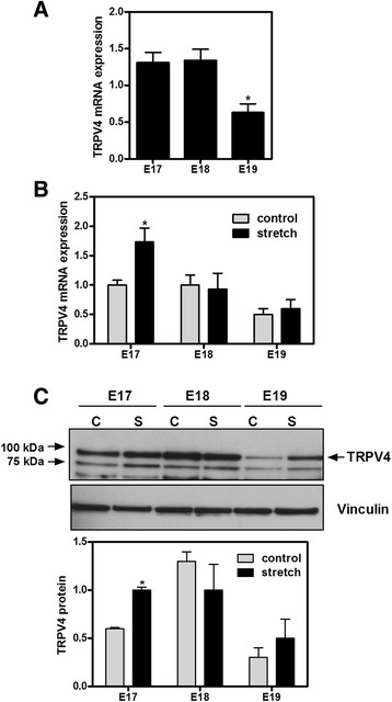
Fig. 2.

TRPV4 expression in isolated distal fetal epithelial cells and the effect of mechanical stretch. a E17-E19 fetal epithelial cells were isolated as described in methods and processed to analyze TRPV4 mRNA expression by qRT-PCR using the ∆∆CT method for relative quantification (n = 4; *p < 0.02 vs E17 or E18, Tukey-Kramer Multiple Comparisons Test). b Fetal epithelial cells were isolated at E17-E19 of gestation and cultured on bioflex plates coated with fibronectin. Twenty four hours later, monolayers were exposed to 20 % cyclic stretch at 40 cycles/min for 24 h; unstretched samples were used as control. Samples were processed by qRT-PCR to assess TRPV4 mRNA expression (n = 4; *p < 0.05 vs E17 control, Tukey-Kramer Multiple Comparisons Test). c Fetal epithelial cells isolated from E17-E19 lungs were seeded on bioflex plates, as described above, and exposed to 20 % cyclic stretch for 24 h. Proteins were extracted and processed to determine TRPV4 protein abundance. The upper panel is a representative blot normalized to vinculin. Data in the lower panel are from 4 different experiments (*P < 0.01 vs E17 control, Tukey-Kramer Multiple Comparisons Test). C = control; S = stretch
