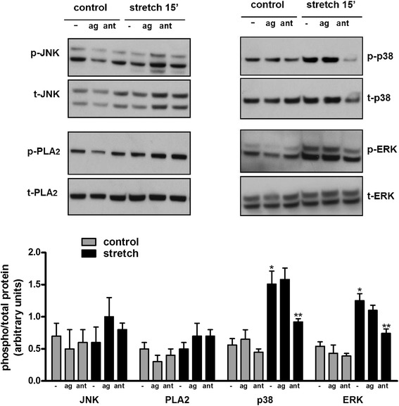
Fig. 4.

Stretch-induced activation of TRPV4 is mediated via p38 and ERK pathways. E17 distal lung epithelial cells were isolated and seeded on plates coated with fibronectin. The following day, monolayers were exposed to 20 % cyclic stretch at 40 cycles/min for 15 min in the presence or absence of the vehicle DMSO, the TRPV4 agonist GSK1016790A [100 nM] or TRPV4 antagonist HC-067047 [1 μM]. The level of activation of the indicated proteins in the cell lysate was evaluated by Western blot using phospho-specific antibodies. Blots were then stripped and reprobed with total antibodies to control for protein loading. Upper panels are representative Western blots. Data in the lower panel are from 5 different experiments. *p < 0.05 vs negative control; **p < 0.01 vs negative stretch. Tukey-Kramer Multiple Comparisons Test
