Figure 1.

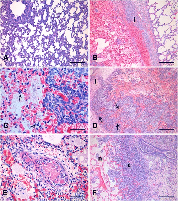
HE-stained lung sections. (A) Lung section from a control pig with mild thickening of the alveolar septa. Scale bar 200 μm; (B-F) Lung sections from pigs inoculated with A. pleuropneumoniae. (B) 6 h p.i.: An interlobular septa [i] with bleeding, oedema and infiltration of neutrophils, separates a non-affected lobule and a lobule with hyperemia, bleeding, oedema, infiltration of neutrophils, bacteria and fibrin. Scale bar 200 μm; (C) 6 h p.i.: Necrosis and degeneration of alveolar septal cells (arrows), and presence of oat-shaped cells [o]. Scale bar 30 μm; (D) 12 h p.i.: Increased cellular infiltration along an interlobular septa [i] and coagulative necrosis of the alveolar septa (arrows). Scale bar 200 μm; (E) 24 h p.i.: Fibrinoid necrosis of a blood vessel. Scale bar 40 μm; (F) 48 h p.i.: Lobule affected by coagulative necrosis [n] enclosed by dense fringes of neutrophils and mononuclear cells [c]. Scale bar 200 μm.
