
Figure 1.
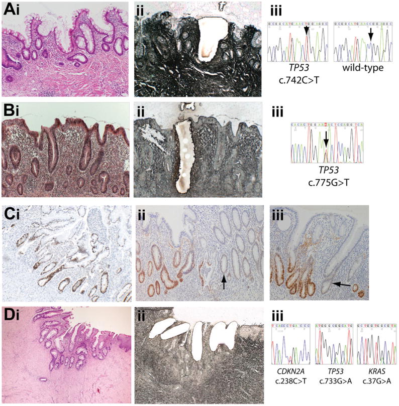
Mutations present in nontumor tissue. (A) (i) H&E stain (original magnification 100×) of nondysplastic (hyperplastic) mucosa showing crypt distortion with increased inflammatory cells and intraepithelial neutrophils in the transverse colon in patient 1 in 2004 and (ii) serial methylene green–stained PALM laser capture slide showing microdissected crypt. (iii) Sequencing shows the crypt contains a TP53 c.742C>T mutation. (B) (i) H&E stain (original magnification 100×) of nondysplastic (hyperplastic) mucosa with a marked increase in the inflammatory cells in the lamina propria and cryptitis in the resection margin of the sigmoid cancer resected from patient 1 in 2000 with (ii) serial PALM slide showing microdissection. (iii) Sequencing shows the crypt contains a TP53 c.775G>T mutation. (C) p53 immunohistochemistry on a nondysplastic colon resection specimen from patient 1 (original magnification 100×). (i) Patches of crypts with nuclear accumulation of p53 protein were observed, frequently demarked by odd p53-negative or low-expressing crypts (ii and iii) (arrows). (D) (i) H&E stain (original magnification 40×) of inflammatory atypia within cells in a perianal fistula from patient 5 four years before tumor growth. (ii) Laser capture slide (original magnification 100×). (iii) TP53, CDKN2A, and KRAS mutations in each crypt.