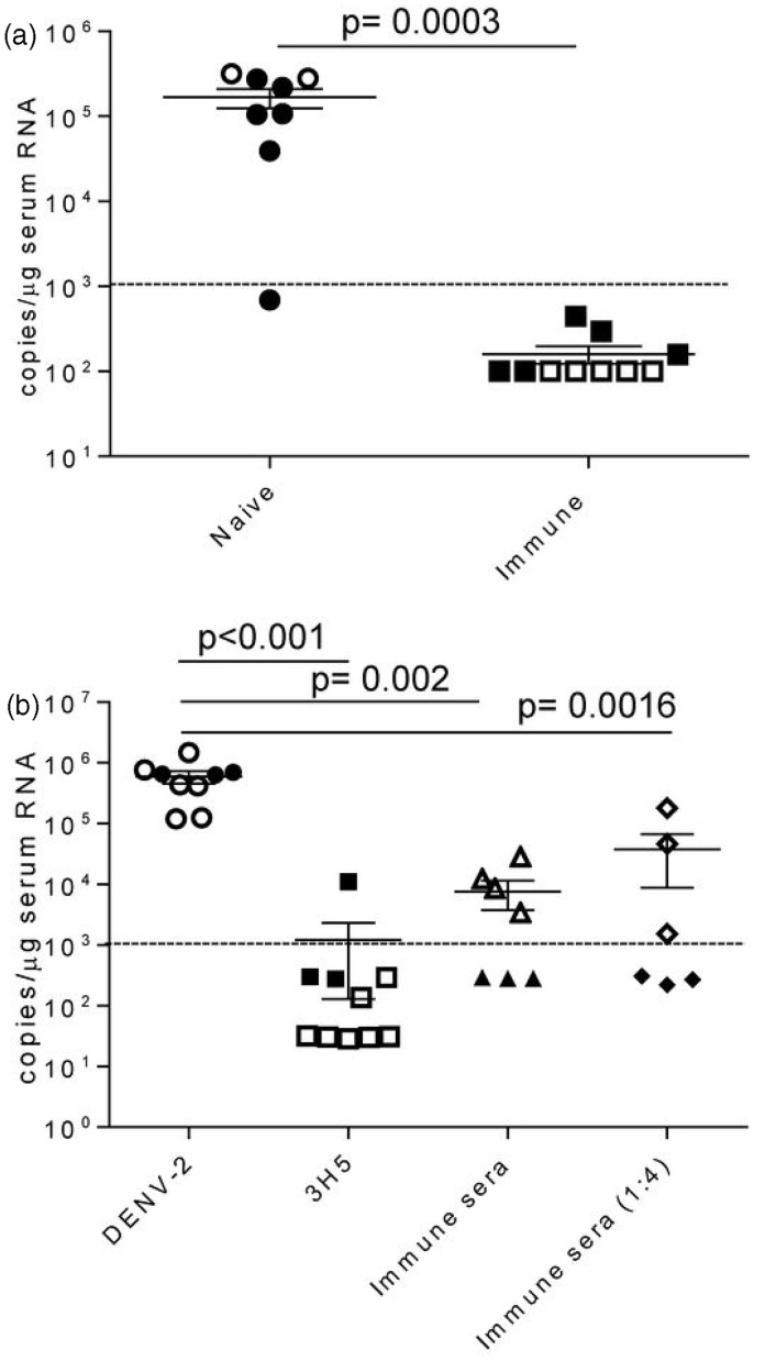
Figure 7.
Decreased viral titers in immune BLT-NSG mice. (a) Mice were immunized three (closed squares) or nine (open squares) weeks prior with 1 × 10⁸ PFU or 6 × 10⁷ PFU of DENV-2 S16803, respectively, by the s.c. route. Matched naïve (closed and open circles) or immunized BLT-NSG mice were inoculated with ∼1 × 10⁶ PFU of a clinical strain DENV-2 C0576/94 by the i.v. route. Mice were bled at day 7and RNA was isolated from serum and subjected to one-step RT-PCR and quantitative PCR. Values represent copy number of viral RNA present per µg serum RNA detected by quantitative RT-PCR. Two separate experiments were performed (open and closed symbols) and were pooled together for analysis; n = 8 naïve BLT NSG and n = 10 immune BLT NSG mice. (b) DENV-2 16681 (1 × 10³ PFU/mL) was incubated with 100 µL PBS (DENV-2), 100 µL 3H5 antibody (3H5), 100 µL BLT-NSG immune serum, and 100 µL diluted (1:4) BLT NSG immune serum for 1 h at 37℃. NSG-Type 1 IFNR KO mice were injected s.c. with 100 µL of a 1:1 mixture and mice were bled at day 5. RNA was isolated from serum and subjected to one-step RT-PCR and quantitative PCR. Values represent copy number of viral RNA present per µg serum RNA detected by quantitative RT-PCR. Two separate experiments were performed (open and closed symbols) and were pooled together for analysis. P values, as determined by Mann Whitney U test, are shown for statistically significant comparisons. Error bars represent the mean with SEM. Dashed line represents the limit of detection

