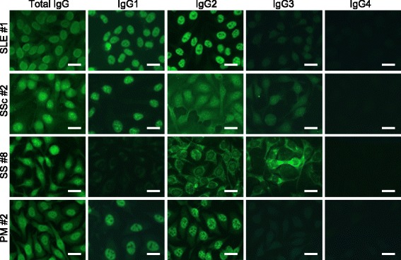
Fig. 3.

Subclass-based ANA test for systemic autoimmune diseases showing immunofluorescence microscopy for each typical case, including systemic lupus erythematosus (SLE #1), systemic sclerosis (SSc #2), Sjögren’s syndrome (SS #8) and polymyositis (PM #2) showed variation in ANA patterns among IgG subclasses. In SSc #2, total IgG showed Discrete spe + Speckled + Cyto, while IgG1 showed Discrete spe + Speckled, IgG2 showed Discrete spe + Speckled + Cyto, IgG3 showed Discrete spe + Cyto, and IgG4 showed negative. In SS #8, total IgG showed Speckled + Nucleolar + Cyto, while IgG1 and IgG2 showed Speckled + Cyto, IgG3 showed Nucleolar + Cyto, and IgG4 showed negative. Bar = 20 μm Discrete spe: discrete speckled, Cyto: cytoplasmic
