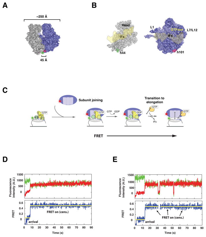
Figure 1. Intersubunit FRET between specifically labeled ribosomal subunits reports on intersubunit dynamics without interfering with ribosome function.
(A) Surface representation of the E. coli 70S ribosome, showing the location of Cy3 (green) and Cy5 (red) labels. The 30S subunit (grey) is labeled at helix 44, whereas the 50S subunit (blue) is labeled at helix 101. (B) View of the 30S (grey) and 50S (blue) subunits from the subunit interface, with an overlay of the estimated binding footprint for the initiation factors (IFs), modeled from structural studies. (C) Surface-immobilization of Cy3-labeled 30S pre-initiation complexes containing 30S subunits (grey), initiation factors (green), tRNA and mRNA, followed by delivery of Cy5-labeled 50S (blue) results in formation of a 70S initiation complex and establishment of a FRET signal sensitive to intersubunit conformation. (D, E) Representative fluorescence vs. time trajectories obtained from single 70S complexes. Raw fluorescence from Cy3 (green) and Cy5 (red) are used to calculate FRET (blue). Upon stop-flow delivery of cy5-50S, an initial dwell time is observed, followed by a burst of FRET. This FRET signal is stable and often censored by the end of observation (D) or less frequently by photophysical events (E).

