
Fig. 1.
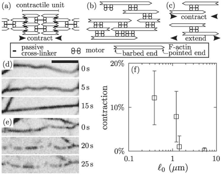
Contraction in actomyosin bundles. (a) Sarcomeric structure as in striated muscle. As motors tend to move toward the filament barbed ends, the sarcomeric structure imposes that each contractile unit (sarcomere) contracts. (b) Bundle devoid of sarcomeric organization or passive cross-linkers, as in our experiments. (c) Motors and polar filaments induce local contraction or extension depending on the geometry of their assembly (filament polarity always dictates the direction of motion [8]). (d) Time-lapse images of a bundle comprised of F-actin and fluorescent myosin thick filaments (inverted contrast) with ℓ0 = 540 nm. The initially wavy bundle becomes taut following the addition of 1 mM ATP at t = 0 s, indicating contraction. Scale bar, 5 μm. (e) Similar experiment with ℓ0 = 1.5 μm, showing no contraction. Scale bar as in (d). See also movie S1 [18]. (f) Bundle contraction as a function of ℓ0. Bars indicate standard deviation (n ≥ 25).