
Figure 1.
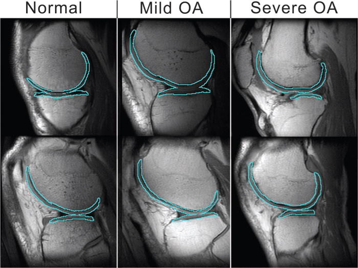
Example of registration between the T2-weighted MR image and the cartilage mask in six subjects (ie, two from each group). Sagittal view of femoral and tibial compartments outlined over the first echo of the dual two-dimensional spin-echo MR image (1,500/10 and 45; voxel size, 0.468 × 0.468 × 4 mm; examination time, 5 minutes 24 seconds; field of view, 12 cm; matrix, 256 × 256). Note that these are only single sections of the entire knee volume and may not reflect specific regions of disease that account for OA grade.