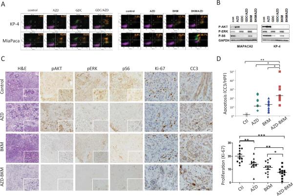
Figure 2. Analysis of cell lines and ex vivo organotypic cultures supports the combined targeting of MEK and PI3K in PDAC.
A, B) Treatment of PDAC cell lines with vehicle control or with the indicated drugs (AZD: AZD-6244; BKM: BKM-120; GDC: GDC-0941) used at 1 μ#. A) FACS plots showing PI/AnnexinV staining. The % of apoptotic cells is indicated. B) Western blot showing effect of the inhibitors on p-AKT (Thr308) and p-ERK (Thr202/Tyr204) levels.
C-D) Analysis of therapeutic responses of ex vivo organotypic cultures of primary PDAC. C) Freshly derived organotypic cultures were treated with the indicated compounds (each at 1 μM) for 24 hours and then processed for staining with H&E or with antibodies to p-ERK (Thr202/Tyr204), p-AKT (Thr308), p-S6 (Ser235/236), Ki-67, and cleaved Caspase 3. D) Quantification of apoptosis at 24 hrs (cleaved caspase-3 staining) and of proliferation (Ki-67 staining). Statistical significance is indicated; p<0.01 (*), p<0.0001 (**).

