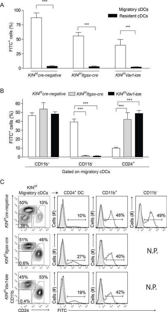
Figure 4. Klf4-dependent CD11b− cDCs account for substantial antigen transport from skin to lymph nodes.
(A) Shown is the contribution to total FITC+ cells by migratory (open bars) and resident (closed bars) cDCs in skin draining lymph nodes (sLN) 16 h after FITC painting, as a percentage of total FITC+ sLN cells. Error bars, ± s.d., n = 6, Student’s t-test. *P < 0.05; ***P < 0.001; NS, P > 0.05. (B) Shown is contribution to total FITC+ cells by each migratory DC subset as a percentage of total FITC+ sLN cells in mice of the indicated genotypes 16 h after FITC painting. (C) FITC transport by individual migratory cDC subsets is show as single color histogram from mice of the indicated genotypes and gates (left panels). N.P. Not Present. Numbers indicate the percent of cells in the indicated gates. The experiment was performed 3 times and 2 mice per group were used. See also Supplemental Figure S4

