
Figure 4.
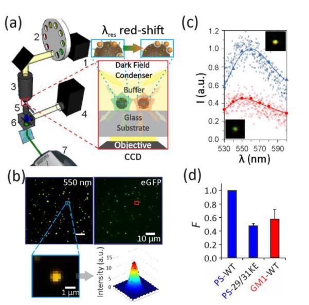
Optical set-up for correlated fluorescence / multispectral darkfield imaging. a) Scheme of the optical set-up to characterize NP labeled VLPs (inset). 1-Tungsten lamp, 2-Filter wheel, 3-Darkfield condenser, 4-Mercury lamp, 5-60x oil objective, 6-Fluorescence filter set, 7-EMCCD. b) Scattering (left) and fluorescence (right) image for WT VLP labeled for PS with NPs. A set of 9 scattering images in the range between 530 to 600 nm were recorded. Inset shows fitted point-spread-function for one scatterer. c) Comparison of spectra obtained through multispectral analysis (continuous lines) and imaging spectrometer (small markers) for an individual VLP with low (red, bottom) and high (blue, top) NP coverage. Insets show the corresponding scattering images. d) The binding probability, F, obtained through three or more optical measurements of three different samples. Error bars indicate standard deviations.