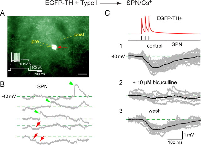
Figure 10.
Paired whole-cell current-clamp recording between a presynaptic Type I neuron and a postsynaptic SPN. Both neurons were recorded with a CsMeSO4 internal solution. A, The Type I neuron (red arrow, pre) is fluorescent and shows a typical response to depolarizing current (inset). The postsynaptic SPN (post) is not fluorescent and cannot be seen under epifluorescence illumination. Scale bar, 20 μm. B, Depolarizing the SPN to −40 mV reveals both spontaneous EPSPs (green arrowheads) and IPSPs (red arrows). C1, Current pulses in the Type I neuron elicit three spikes that evoke three hyperpolarizing IPSPs in the SPN at −40 mV that summate to >1 mV. C2, The IPSP was reversibly blocked by bicuculline (10 μm) and recovered after wash (C3), showing that the synaptic response was mediated by a GABAA receptor.

