
Figure 11.
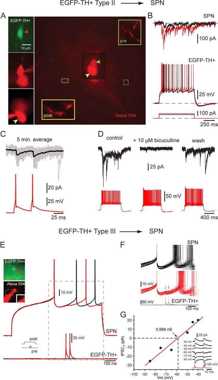
Spiking in Type II and Type III neurons elicits IPSCs in SPNs and delays firing in response to current injection. A, Connected pair consisting of a presynaptic EFGP+ Type II neuron (top left) and a postsynaptic SPN (bottom left). The insets are high-magnification micrographs showing an aspiny Type II (pre) dendrite and a spiny SPN dendrite (post) after both were patched in whole-cell mode and stained with Alexa 594. B, Top, Current pulses injected into the Type II neuron elicited a train of presynaptic spikes that evoked IPSCs in the postsynaptic SPN. Both short-term depression and early facilitation were evident. C, Two spikes evoked by two 5 ms pulses 50 ms apart in the Type II neuron result in modest paired-pulse facilitation in the same SPN. Black traces are averages, and gray traces are individual trials. D, The IPSCs were reversibly blocked by bicuculline (10 μm) showing that they were mediated by a GABAA receptor. E, Connected pair consisting of a presynaptic Type III neuron and a postsynaptic SPN (inset). Two presynaptic spikes separated by 50 ms (bottom trace) delay the occurrence of depolarization induced spiking in the postsynaptic SPN (top black trace) by almost 100 ms (top red trace). F, Entire dataset from which the traces in E were selected. Depolarization evoked spiking in SPN under control conditions (black traces) and in the presence of two spikes in the presynaptic neuron (red traces). Note the reliability of this connection. G, I–V plot for this synaptic response showing a reversal potential consistent with mediation by Cl− ions and a synaptic conductance near 1 nS.