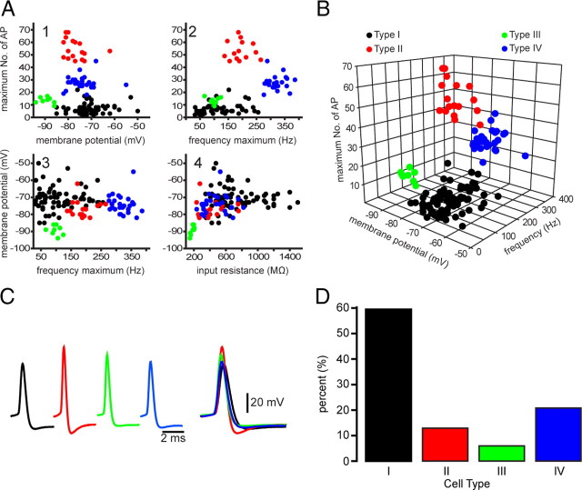
Figure 2.
Four different types of EGFP–TH+ neurons in mouse striatum. A, Selected two-dimensional scatter plots of various electrophysiological parameters reveal the separation of striatal EGFP–TH+ neurons into four distinct groups, termed Types I–IV. AP, Action potential. B, Clustering of four distinct cell types in one representative three-dimensional scatter plot. C, Averaged action potentials from cell Types I–IV clearly show differences in multiple spike waveform parameters. D, Histogram showing the distribution of the four EGFP–TH+ cell types.

