
Figure 3.
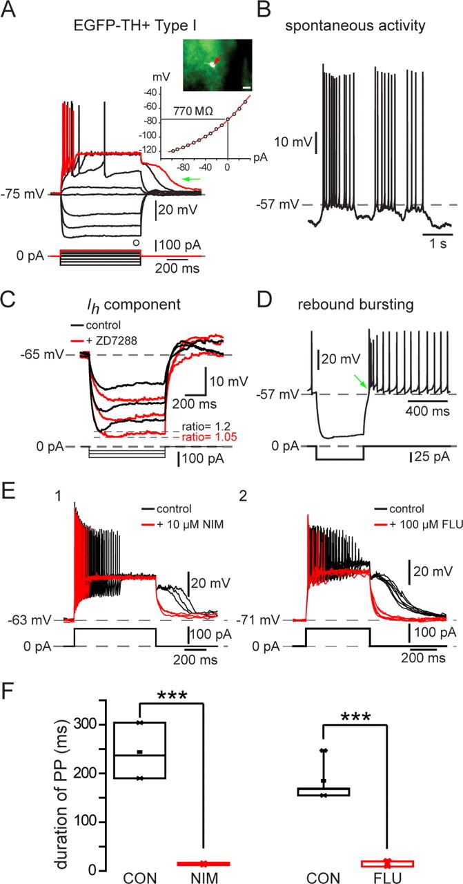
Electrophysiological properties of Type I EGFP–TH+ neurons. A, Whole-cell current-clamp recordings of responses to negative and positive current pulses in a Type I neuron show high input resistance, strong spike frequency adaptation leading to complete spike failure after small depolarizing pulses (red trace), and the expression of long-lasting plateau potentials (green arrow). Inset, EGFP fluorescent image of recorded neuron and I–V plot showing inward rectification. I–V curves in this and Figures 4–6 are plotted at the end of the current pulse at the point marked by an open circle. B, Some Type I neurons exhibited slow, irregular spontaneous activity characterized by ∼10 mV fluctuations in membrane potential. C, Time-dependent sag in response to hyperpolarizing current injection and rebound slow depolarization after its offset (green arrow) are both blocked by ZD7288 (50 μm), indicating that both are attributable to Ih. D, Rebound bursting after offset of hyperpolarizing current injections. E1, The plateau potential was blocked by 10 μm nimodipine (NIM). E2, Plateau potential in a different Type I neuron is blocked by 100 μm flufenamic acid (FLU), indicating that the plateau potentials are attributable to a Ca2+-activated cation conductance (ICAN). F1, Summary of effects of nimodipine on plateau potential (PP) duration in six Type I neurons. F2, Summary of effects of flufenamic acid on plateau potential (PP) duration in eight Type I neurons. CON, Control. ***p < 0.0001.