
Figure 4.
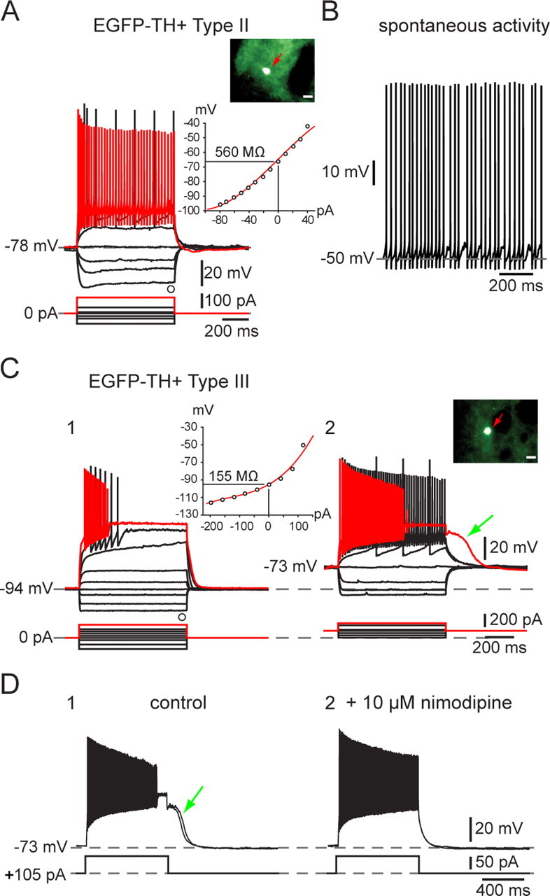
Electrophysiological properties of Type I and Type II EGFP–TH+ neurons. A, Responses to negative and positive current pulses in a Type II neuron illustrate lower input resistance than Type I neurons, similar time-dependent Ih-like sag in response to hyperpolarizing current injections, and ability to sustain high-frequency firing with little spike frequency adaptation after the first few milliseconds. Inset, EGFP fluorescent image of recorded neuron and I–V plot showing almost linear responses except at most hyperpolarized membrane potentials. B, Example of tonic spontaneous firing. C, D, Electrophysiological properties of Type III EGFP–TH+ neurons. C1, Responses to negative and positive current pulses in a Type III neuron reveal a linear I–V relationship below −90 mV and marked inward rectification at more depolarized potentials. Note the very low input resistance and the very hyperpolarized resting membrane potential compared with Type I and Type II neurons. There is significant spike frequency adaptation, leading to complete spike failure in responses to strongest depolarizing current pulses. C2, When Type III neurons are depolarized by constant current injection, overall excitability is increased, spike frequency adaptation is reduced allowing sustained firing up to 120 Hz, and a prolonged plateau potential appears (green arrow). The plateau potential (D1) is blocked by 10 μm nimodipine (D2), demonstrating the involvement of an L-type Ca2+ channel.