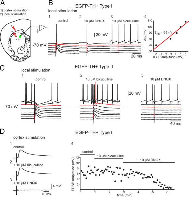
Figure 9.
Synaptic input to EGFP–TH+ neurons. A, Experimental design. Bipolar stimulating electrodes were placed onto the surface of the deep layers of the cortex (1) and in striatum to evoke responses in striatal EGFP–TH+ neurons. CPu, Caudate putamen. B1, In a representative Type I cell, local stimulation evoked a biphasic response consisting of an overlapping EPSP and IPSP (control). B2, DNQX (10 μm) blocked the EPSP component, leaving a pure IPSP with an apparent reversal potential of −65 mV (B4). B3, The IPSP was blocked by bicuculline (10 μm), indicating mediation by GABAA receptors. C, Same experiment performed with a Type II neuron but reversing the order of the antagonist application. C1, Local stimulation evokes biphasic response. C2, The inhibitory component was blocked by 10 μm bicuculline, showing that it was mediated by a GABAA receptor. C3, The remaining EPSP is completely eliminated by addition of DNQX (10 μm). D1, Cortical stimulation evokes short-latency monosynaptic DPSP in a Type I neuron. D2, The DPSP is not affected by 10 μm bicuculline. D3, The AMPA/kainate channel blocker DNQX (10 μm) completely eliminates the DPSP, showing that it is a glutamatergic EPSP. D4, Time course of drug effects on the EPSP.

