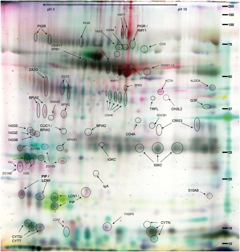
Fig 2. False colour two-dimensional differential gel electrophoresis (2D-DIGE) image of identified protein spots in sputum.
False colour image of the identified protein spots (circled and named) on a 2-D DIGE-gel of the sputum fluid phase samples of 21 subjects subdivided as follows: asthma with allergic rhinitis, allergic rhinitis, nonallergic rhinitis, and healthy controls. Samples of the subjects with asthma with allergic rhinitis (reddish up-regulated) and with nonallergic rhinitis (green up-regulated), as well as the internal standard are shown. The identified proteins are listed in S1 Table.

