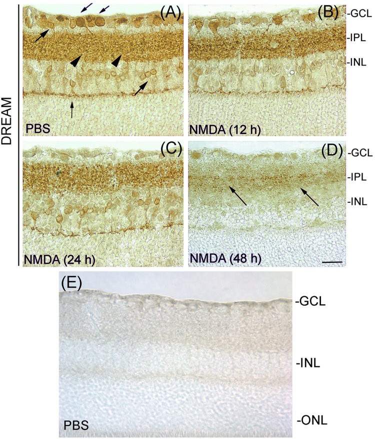
Fig 1. Expression of DREAM in the retina.
Retinal cross sections prepared after treating the eyes with PBS or NMDA were immunostained with antibody against DREAM. Results presented in panel A show that DREAM was expressed in RGCs (down word arrows), amacrine cells (up word arrows), bipolar cells (vertical arrow), as well as in the IPL (arrow heads) in retinal cross sections prepared from PBS-treated eyes. DREAM expression was decreased following NMDA-treatment (panels B and C), and at 48 h after the treatment, very low DREAM levels were detectable in the IPL (panel D). Panel E, negative isotype control. Bar indicates 50 microns size.

