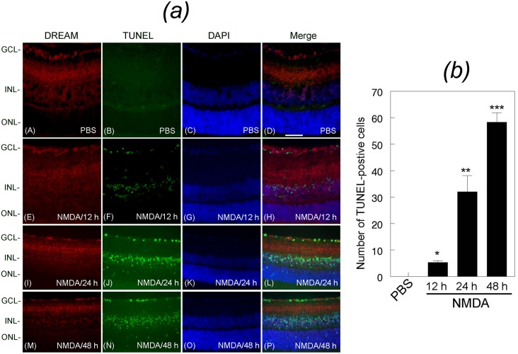
Fig 3. Expression of DREAM and apoptotic cell death in the retina.
(a) Retinal cross sections prepared from PBS or NMDA-treated eyes were immunostained with antibody against DREAM (panels A, E, I, and M), subjected to TUNEL assays (panels B, F, J, and N), counter stained with DAPI (panels C, G, K, and O), and the images were merged (D, H, L, and P). Results presented in the figure show that DREAM was expressed in the GCL, IPL, and INL in retinal cross sections prepared from PBS-treated eyes (panels A and D). DREAM expression was decreased at 12 h (panels E and H), 24 h (panels I and L), and 48 h (panels M and P) after NMDA treatment. No TUNEL-positive cells were observed in retinal cross sections prepared from PBS-treated eyes (panel B), but increased number of TUNEL-positive cells were observed in retinal cross sections prepared from NMDA-treated eyes (panels F, J, N). Merged images indicate that TUNEL-positive cells were localized initially in the GCL (panel H), and later in the INL (panel L) and the ONL (panel P). Bar indicates 50 microns size. (b) Quantitative analysis indicates that when compared to PBS treatment, NMDA treatment promoted apoptotic cell death significantly over a 48 h time period,.*, **, *** p<0.05.

