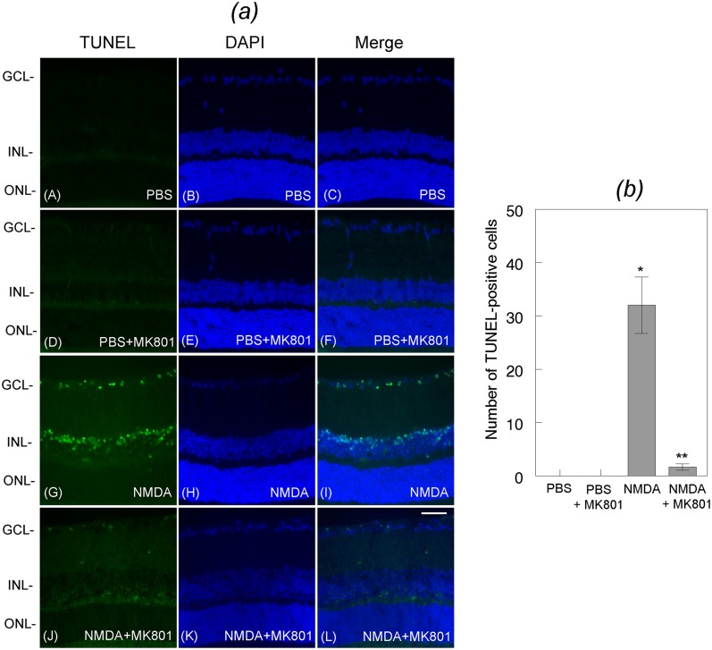
Fig 8. Effect of MK801 on NMDA-mediated apoptotic death of retinal neurons.
Retinal cross sections prepared from PBS or NMDA-treated eyes were subjected to TUNEL assays. Results presented in the figure indicate that when compared to PBS [Fig. (a), panels A, B, C], NMDA promoted significant apoptotic cell death in the GCL and the INL at 24 h after the treatment [Fig. (a), panels G, H, I]. In contrast, treatment of the eyes with NMDA and MK801 significantly attenuated NMDA-mediated apoptotic death of cells both in the GCL and INL [Fig. (b)]. Bar indicates 50 microns size. Characterization of neuronal cell’ loss in whole retinas and in retinal cross sections indicates that NMDA promoted a significant loss (*p<0.05) of Brn3a-positive RGCs (C), calretinin-positive amacrine cells (D), and PKC-alpha-positive bipolar cells (E), and MK801 attenuated the loss of all three cell types (**p<0.05).

