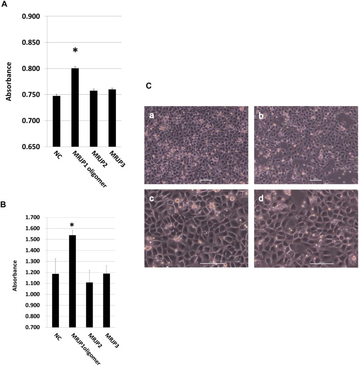
Fig 6. Cell proliferation activity of MRJP1 oligomer, MRJP2 and MRJP3.
(A) Cell proliferation activity on Jurkat cells. Cells were cultured with DME/F-12 medium containing 0.5 mg/ml each protein for 24 hours. For negative control (NC), cells were cultured with DME/F-12 medium without any proteins. Cell proliferation was measured by Alamar Blue assay. Error bar indicate S.E.M. from three independent experiments (n = 3). Asterisk indicates p<0.01 vs NC. (B) Cell proliferation activity on IEC-6 cells. Cells were cultured in D-MEM medium containing 0.5 mg/ml each protein for 24 hours. For negative control (NC), cells were cultured with D-MEM medium without any proteins. Cell proliferation was measured by WST-8 assay. Error bar indicates S.E.M. from three independent experiments (n = 3). Asterisk indicates p<0.05 vs NC. (C) Phase-contrast images of IEC-6 cells. Cells were cultured in D-MEM medium containing 0.5 mg/ml MRJP1 (a, c) and without any protein (b, d) for 24 hours. Scale bar indicates 100μm. Magnification of figure a, b and figure c, d are 100-fold and 200-fold, respectively.

