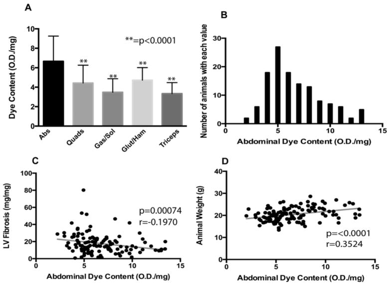
Figure 2. Mean dye uptake, as marker of muscle damage, was highest in the abdominal muscles.
A large cohort (n=196) of SgcgD2/129 mice was assayed for dye uptake in multiple muscle groups. A) Mean dye uptake was found to be highest in the abdominal muscles (m=6.67 ± 2.59 OD/mg), This value was found to be significantly higher than other muscles assayed for EBD when compared by Dunn’s multiple comparison tests (p<0.0001). Error bars represent standard deviation. B) A histogram representing the distribution of abdominal muscle dye uptake. This distribution was found to be non-normal, and right-skewed. C) Abdominal dye uptake was significantly, negatively correlated with fibrosis in the left ventricle of the heart. D) Furthermore, abdominal dye uptake was significantly, positively correlated with animal weight. Taken together, these data support a model in which abdominal muscles are recruited to support cardiopulmonary output.

