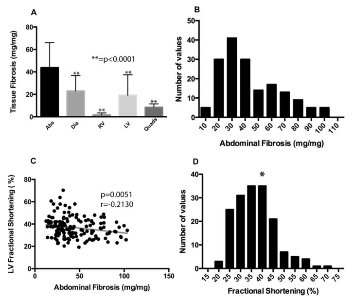
Figure 3. Mean tissue fibrosis was highest in the abdominal muscles.
SgcgD2/129 mice from the same cohort (n=196) were assayed for hydroxyproline (HOP) content as a direct measure of fibrosis. A) Mean fibrosis, measured by HOP content, was highest in the abdominal muscles (43.79 ± 22.16 mg/mg). This value was found to be significantly higher than other muscles when compared by Dunn’s multiple comparison tests (p<0.0001). Error bars represent standard deviation. B) A histogram representing the distribution of abdominal muscle fibrosis (HOP content.) This is a non-normal distribution and is right-skewed. C) Abdominal muscle fibrosis was significantly negatively correlated with left-ventricular fractional shortening (LVFS) using Spearman based calculations. D) Distribution of LVFS in the Sgcg cohort. The asterisk indicates normal LVFS.

