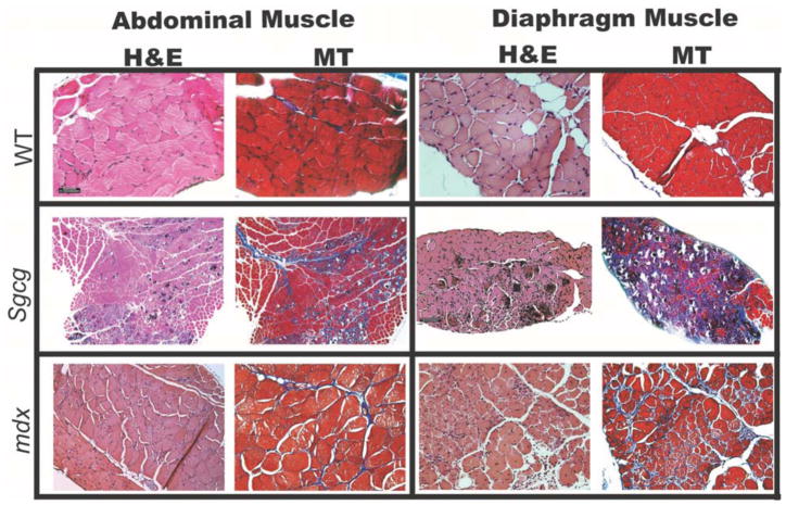
Figure 5. Abdominal muscle and diaphragm muscle histopathology is comparable in mouse models of MD.
Representative histopathologic sections taken from the diaphragm and abdominal muscles of normal wild-type (WT), Sgcg, and mdx and Sgcg mice at an early time point in disease. All mice were less than 12 weeks old. Hematoxylin and eosin (HE) staining highlights necrosis, calcification, and inflammation. Mason’s trichrome (MT) stained sections from Sgcg and mdx mice both revealed a more dense intercellular fibrosis throughout the abdominal and diaphragm muscles. This degree of histopathology is greater than what is seen in limb-based skeletal muscles (not shown).

