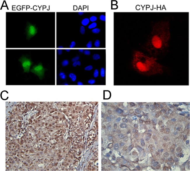
Fig 3. Subcellular localization of CYPJ.

(A) Hela cells were transfected with pEGFP-CYPJ. After 48 h, cells were fixed and stained with DAPI to indicate the nuclei. (B) Hela cells were transfected with pCMV-CYPJ-HA, and 48 h later, the cells were fixed and stained with anti-HA monoclonal antibody. (C) and (D) Immunohistochemical stain of human HCC tissue section using anti-PPIL3 antibody. (C) ×100 magnification. (D) ×400 magnification.
