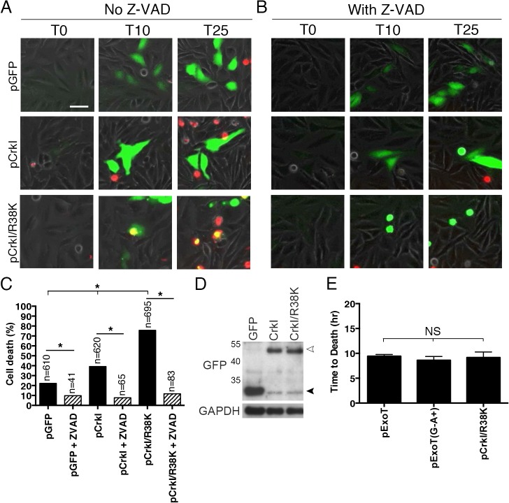
Fig 4. CrkI/R38K mutant phenocopies ExoT/ADPRT-induced apoptosis in HeLa cells.
HeLa cells were transiently transfected with pIRES2-GFP expression vector harboring wild type CrkI (pCrkI), SH2 DN (pCrkI/R38K), all directly fused to GFP C-terminally, or empty vector (pGFP) in the absence (A) or presence (B) of Z-VAD pan-caspase inhibitor. PI was added to identify dying cells and cell death was analyzed by timelapse IF videomicroscopy. (C) The tabulated results, collected from multiple movies, are shown. (D) Transfection efficiencies were evaluated by Western blotting, using anti-GFP antibody and cells lysates from HeLa cells transfected as in (A). GAPDH was used as a loading control. The black arrowhead points to the position of GFP from the vector alone and the white arrowhead indicates CrkI-GFP and CrkI/R38K-GFP. (E) The time to death was defined as the time of expression of the transfected gene (appearance of green) to the time of PI uptake (appearance of yellow) and expressed as the mean ± SEM. Note that expression of SH2 DN CrkI induces potent apoptosis and kinetically phenocopies ExoT and ExoT/ADPRT-induced apoptosis. (* Signifies significance with p<0.01, χ2 analyses. Scale bar = 25 μm).

