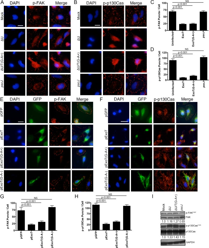
Fig 7. ExoT and ADPRT disrupt focal adhesion sites.
(A-D) HeLa cells were treated with PBS or infected with ∆U, ∆U/T(G-A+), or pscJ at MOI~10. Four hours after infection, cells were fixed and stained with nuclear stain DAPI and p-FAK (A) or p-p130Cas (B). Representative images are shown in (A and B) and the tabulated results are shown in (C and D). (E-H) HeLa cells were transiently transfected with pIRES2-GFP expression vector harboring ExoT (pExoT), ExoT/ADPRT (pExoT(G-A+), or inactive ExoT (pExoT(G-A-), all C-terminally fused to GFP, or empty vector (pGFP). 24 hr after transfection, cells were fixed and stained with nuclear stain DAPI and analyzed for p-FAK and p-p130Cas localization to the FA sites. Representative images are shown in (E and F) and the tabulated results from 3 independent experiments are shown in (G and H). (Statistical analysis was performed using one-way ANOVA. Scale bar = 25 μm). (I) HeLa cells were infected as in (A). 4hr after infection, the cell lysates were probed for total FAK and p130Cas or their phosphorylated forms (p-FAKY397 & p-p130CasY165 respectively) by Western blotting. Expression levels were normalized to GAPDH, which was used as the loading control.

