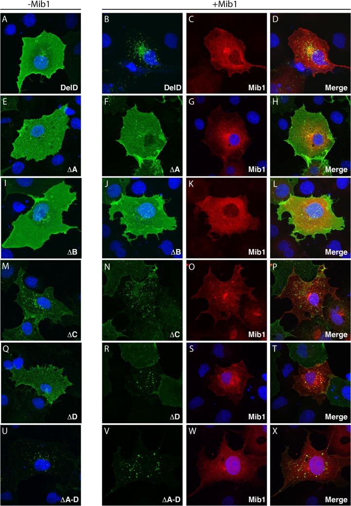
Fig 3. Endocytosis of DeltaD deletion mutants.
(A,E,I,M,Q,U) Distribution of zdd2 (green) in COS7 cells transfected with DeltaD (A) or DeltaD ∆A, ∆B, ∆C, ∆D or ∆A-D deletion mutants (E, I, M, Q, U). Surface DeltaD was first labelled by incubation with zdd2 at 4°C for 30’ then, following washout of unbound zdd2, internalization was allowed for 30’ at 37°C. Nuclei were labelled with DAPI (blue). (B-D, F-H, J-L, N-P, R-T, V-X) Distribution of zdd2 (green) in COS7 cells co-transfected with DeltaD constructs and Mib1 (red) following internalization as described above. Each set of 3 panels, respectively, shows distribution of the DeltaD construct (green), Myc-Mib1 (red)/nuclei (blue), and the merged image. See materials and methods for details.

